How digital scanning transforms the patient experience
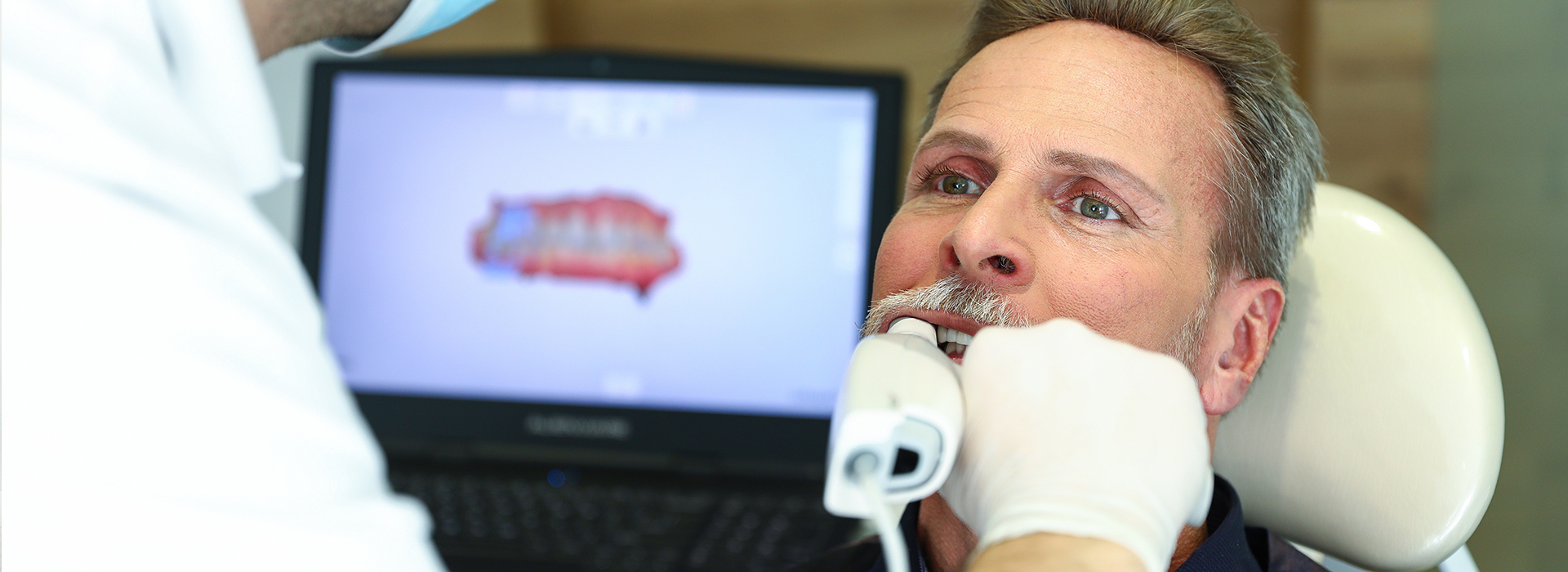
For many patients, the idea of a dental impression brings to mind trays of goo and a gagging reflex. Digital impressions replace that process with a small, handheld intraoral scanner that captures detailed images without bulky materials. The scanner sweeps across the teeth and surrounding tissues while software stitches the data into a precise three-dimensional model, making the visit more comfortable and less stressful for people of all ages.
Beyond comfort, the noninvasive nature of the scan means fewer retakes and less chair time. Because the clinician can review the image in real time, any areas that need extra detail are rescanned immediately, eliminating the uncertainty that sometimes follows traditional impressions. This responsiveness reduces scheduling delays and helps patients move more quickly from diagnosis to treatment planning.
Patients who are anxious about dental procedures often appreciate the predictable, streamlined workflow that digital scanning enables. The procedure is quiet, fast, and does not require setting materials to harden in the mouth. For anyone who struggles with strong gag reflexes, limited mouth opening, or heightened sensitivity, modern digital impressions present a markedly improved experience.
Precision and predictability: what modern scans capture
Intraoral scanners produce highly accurate digital models that record not only the shape of each tooth but also surrounding soft tissues, margin detail, and bite relationships. This level of fidelity allows clinicians and dental laboratories to design restorations that fit more closely to the prepared tooth, reducing the need for manual adjustments during try-in appointments. The result is a restoration that looks and functions as intended from the outset.
Digital files capture the spatial relationships between opposing arches and record occlusal contacts with clarity. That information is invaluable for creating crowns, bridges, and implant restorations where proper bite alignment is critical. When a laboratory or a CAD/CAM system receives a complete, accurate data set, the risk of miscommunication is minimized and the predictability of the final restoration improves.
Another advantage of digital capture is consistency. The scanning workflow is standardized and repeatable, which leads to more predictable outcomes across patients and cases. Clinicians can archive and compare scans over time, monitoring changes and planning treatments with a documented baseline rather than relying solely on memory or physical models.
Speed, communication, and seamless lab integration
One of the most tangible benefits of digital impressions is the speed with which information can be shared. Once a scan is complete, the digital file can be transmitted electronically to a dental laboratory or an in-house design system within minutes. This eliminates shipping delays and reduces the chances of material distortion that can occur with traditional impressions and stone models.
Digital workflows also foster clearer communication. Annotated scans, sectional views, and precise digital margins allow dental technicians to understand the clinician’s intentions without interpreting a physical impression or handwritten notes. Many labs now accept common file formats (such as STL) and work directly with designers to refine contours, contacts, and esthetic details before fabrication begins.
Because digital scans are files rather than fragile models, they can be backed up, duplicated, and shared with specialists as needed. This interoperability supports multidisciplinary care — for example, when coordinating an implant case with a surgeon or when planning a complex restorative sequence that involves multiple providers.
Quality control also benefits from the digital approach: lab technicians and clinicians can review scans together and flag any areas requiring refinement before fabrication. This collaborative review reduces remakes and promotes a smoother path to final restoration.
Same-day restorations: how in-office workflows work
Digital impressions are a cornerstone of same-day dentistry. When combined with in-office CAD/CAM milling and high-strength ceramic materials, a single appointment can include tooth preparation, digital capture, restoration design, and placement. For many patients this means fewer visits and a faster return to normal function.
During the same-day workflow, the scanned data are used to design the restoration on integrated software. The design is then sent to a milling unit or a lab for fabrication. Modern ceramics and bonding protocols enable durable, esthetic results that are suitable for many single-unit crowns and onlays. Clinicians determine case suitability based on clinical factors, and when indicated, patients benefit from an efficient and tightly coordinated process.
Even when same-day fabrication is not appropriate, digital impressions still accelerate the overall course of treatment. The improved accuracy and rapid transmission to external labs shorten turnaround times for crowns, bridges, and implant prosthetics, which means patients spend less time waiting for their definitive restorations.
Importantly, the in-office digital workflow remains rooted in careful clinical judgment. Successful same-day care depends on proper case selection, material choice, and precise scanning technique to ensure long-term function and esthetics.
What to expect during your digital impression appointment
Before your appointment, there is typically no special preparation required beyond routine oral hygiene. During the visit, your clinician will explain the scanning process and prepare the area to be scanned by cleaning and isolating the teeth as needed. A small wand-like scanner is then moved over the surfaces to capture overlapping images that the software assembles into a complete model.
A typical full-arch scan can take only a few minutes, while a targeted scan for a single tooth or area may take even less time. Many patients find the experience quick and comfortable, with no unpleasant tastes or hardening materials in the mouth. If additional detail is needed, the clinician will rescan localized areas on the spot to ensure completeness and accuracy.
After scanning, the clinician reviews the digital model with the patient, pointing out margins, neighboring teeth, and occlusal contacts when relevant. This immediate visual feedback can help patients better understand their diagnosis and the planned restoration. The digital record is then archived in the patient’s chart and used for communication with the laboratory or for in-office design and milling.
Privacy and data security are important considerations for any digital workflow. Dental practices follow professional standards for record keeping and secure transmission of digital files so that patient data are handled responsibly throughout the treatment process.
Digital impressions represent a practical improvement in accuracy, comfort, and efficiency for modern restorative dentistry. The office of Cruzin' Dental uses current scanning technologies to streamline treatment planning, improve lab communication, and support same-day options when clinically appropriate. If you’d like to learn more about how digital impressions could benefit your care, please contact us for more information.


