An intraoral camera is a compact, pen-sized imaging tool that captures detailed, full-color pictures from inside the mouth and displays them instantly on a monitor. These high-resolution images give both clinicians and patients a close-up look at teeth, gums, and other oral structures that are difficult to see with the naked eye. Widely used in modern dental practices, the intraoral camera enhances diagnosis, documentation, and patient understanding without adding complexity to the appointment.
How the intraoral camera captures a clearer picture of oral health
Inside the slim housing of an intraoral camera are powerful lenses, LED lighting, and a digital sensor that work together to produce crisp images even in tight or dark spaces. The device is designed to be maneuverable, allowing dentists and hygienists to angle the lens around molars, between teeth, and along the gum line. Because the lighting is built into the tip, images are consistently illuminated and free from the shadows that can obscure traditional visual exams.
Images are transmitted in real time to a chairside display, which means the dentist can freeze, zoom and annotate photos on the spot. This immediacy helps with accurate assessment of small fractures, worn restorations, early decay or soft-tissue concerns that might not be obvious during routine inspection. The clarity of the images also reduces guesswork, helping clinicians confirm findings before recommending next steps.
Intraoral cameras come in a range of formats — wired or wireless, single-use sheaths for infection control, and with variable focal lengths for close-up or wider views. Despite these technical options, the practical result is the same: better visual information, collected quickly and noninvasively, that supports precise clinical decisions and improved patient communication.
Why patients benefit: seeing is understanding
For many patients, the difference between a verbal explanation and a clear, magnified image is dramatic. When an intraoral photograph appears on the screen, it provides an objective visual reference that helps people understand what the dentist is describing. This reduces uncertainty and makes it easier to follow recommendations because patients can see the issue rather than just hearing about it.
Beyond immediate explanation, images captured during the visit can be saved and reviewed over time. This longitudinal view lets patients and clinicians track changes, measure progression, and confirm whether preventive measures or treatments are working. When patients participate in that visual review, they tend to feel more informed and engaged in their oral health plan.
Because the camera is gentle and quick to use, capturing images adds little to the exam experience. It can also be an especially helpful tool for families, allowing parents to see pediatric concerns, and for patients who prefer to learn visually rather than through technical descriptions. Clear visuals empower patients to ask focused questions and make well-informed decisions about their care.
Clinical advantages: detection, documentation, and planning
In clinical practice, intraoral cameras play multiple roles. They help dentists spot early signs of decay, hairline cracks in teeth, leaky margins around restorations, and localized gum inflammation. Detecting these issues sooner often leads to more conservative and effective treatment options. In cases where a lesion or irregularity is present, a high-quality photograph can be invaluable in guiding the next steps of care.
Documentation is another core benefit. Images are stored in the patient record alongside radiographs and clinical notes, creating a visual timeline of oral health. These records improve continuity of care — both within the practice and when collaborating with other dental specialists or laboratories. A clear photo can simplify communication about the exact location and nature of a problem, reducing misunderstandings.
For treatment planning, intraoral images support accurate restoration design and verification. Dentists can reference images when discussing material choices, the extent of repair needed, or the positioning of crowns and bridges. In restorative and cosmetic workflows, photographs help ensure the final outcome aligns with the clinical plan discussed with the patient.
How intraoral imaging fits into a digital dental workflow
Modern dentistry increasingly relies on integrated digital systems, and intraoral cameras are a natural complement to digital radiography, CBCT scans, and digital impressions. Images captured chairside can be exported into practice management software or digital case files, making them available for treatment planning, insurance documentation, or lab communication. This interoperability streamlines clinical operations and preserves high-quality visuals across the care continuum.
When collaborating with specialists — for example, an endodontist, periodontist, or prosthodontist — sharing clear intraoral images can accelerate referrals and provide additional context that enhances remote case review. Laboratories can benefit from annotated photos when fabricating restorations, ensuring color, shape and margin details are understood before work begins. These efficiencies save clinical time while reducing the likelihood of remakes or adjustments.
From a record-keeping perspective, digital images improve auditability and clinical transparency. They provide objective evidence of conditions observed during appointments, which supports clinical notes and can be referenced if questions arise later. By combining photographs with other diagnostic data, dental teams can build a comprehensive and accessible patient history.
What patients can expect during an intraoral camera exam
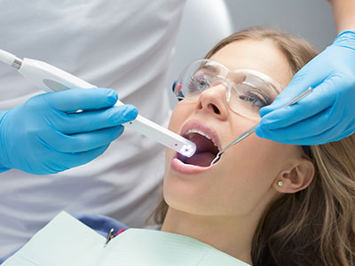
Using an intraoral camera is typically quick and comfortable. The device is small and maneuvered gently into the mouth while the patient is seated comfortably. Because the camera’s tip is smooth and often covered with a disposable sheath for infection control, the experience is similar to routine dental examination instruments — noninvasive and unobtrusive.
The clinician will position the camera to capture the areas of interest and may ask the patient to open, close, or shift their head slightly to obtain the best angle. Images appear on a screen immediately, and the dentist or hygienist can point out features and explain what the photographs show. This live demonstration helps patients understand the nature and significance of any findings as they occur.
After the visit, select images can be appended to the patient’s record for future comparison. If further treatment is recommended, the clinician will use the images to explain the reasoning and outline next steps. Overall, intraoral imaging enhances clarity and supports collaborative decision-making without altering the routine of a standard dental exam.
At Cruzin' Dental, we integrate intraoral imaging into our examinations to improve diagnosis and help patients better understand their oral health. If you’d like to learn more about how this technology may be used during your visit, please contact us for more information.